CIS Conversion Risk Calculator
Calculate Your Risk
Estimate the 5-year conversion risk from Clinically Isolated Syndrome (CIS) to Multiple Sclerosis (MS) based on MRI findings.
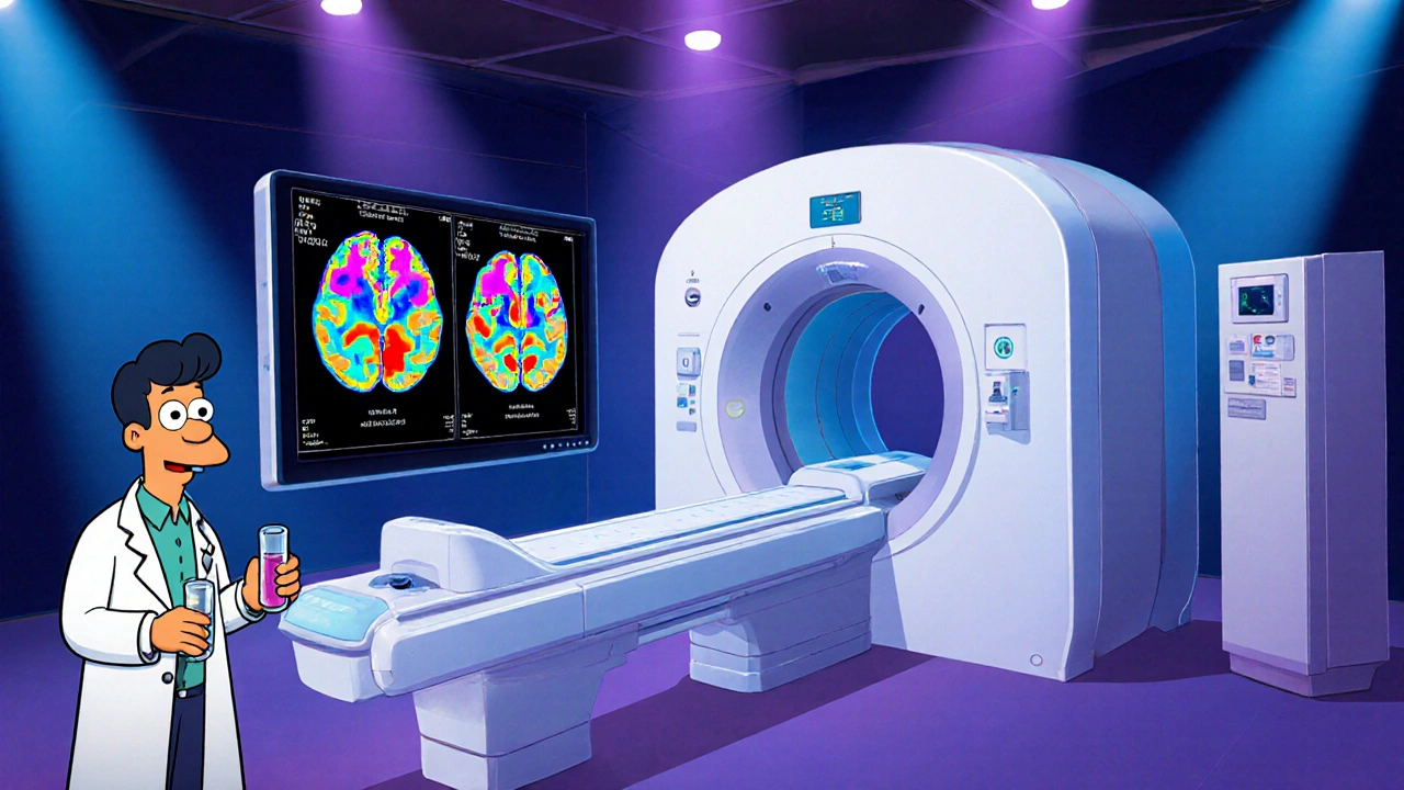
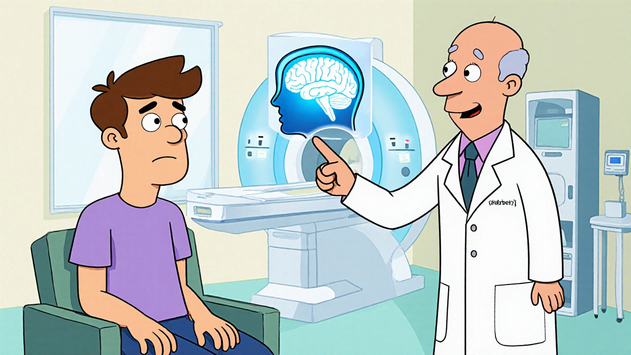
When a person experiences a sudden neurological event-like optic neuritis or a brief limb weakness-and magnetic resonance imaging (MRI) shows a single lesion, doctors label it Clinically Isolated Syndrome (CIS). It’s a warning sign that the immune system may be attacking the brain’s myelin, but it’s not yet full‑blown multiple sclerosis (MS). Catching CIS early can change the whole trajectory, because the window between the first symptom and a possible conversion to MS is the most treatable period.
What Exactly Is Clinically Isolated Syndrome?
Clinically Isolated Syndrome is defined as a single episode of neurologic dysfunction lasting at least 24 hours, accompanied by MRI evidence of demyelination in the central nervous system. Unlike established MS, CIS does not require multiple attacks or disseminated lesions in time and space. About 30-40% of people with CIS will develop MS within five years, but the risk shoots up to 70% when certain MRI features are present.
Why early detection Makes a Difference
Early detection isn’t just a buzzword; it’s a clinical lever. Studies from the MSBase registry (2023) show that initiating disease‑modifying therapy (DMT) within six months of a CIS event reduces the 2‑year conversion rate from 45% to roughly 20%. The reason is simple: DMTs dampen the autoimmune attack before irreversible nerve damage accumulates.
Beyond statistics, early identification gives patients psychological certainty. Knowing the risk empowers them to make lifestyle adjustments-like vitamin D optimization, smoking cessation, and regular exercise-that further lower conversion odds.
Core Diagnostic Tools: Beyond the First MRI
- Magnetic Resonance Imaging (MRI) - the gold standard. High‑resolution T2‑weighted and FLAIR sequences reveal lesions that are invisible on CT.
- CSF oligoclonal bands - presence indicates intrathecal IgG synthesis, a hallmark of MS pathology.
- Evoked potentials - measure the speed of electrical signals through the visual, auditory, or somatosensory pathways; delayed conduction supports demyelination.
- Blood tests for vitamin D, B12, and autoimmune markers help rule out mimics.
Each test adds a piece to the puzzle. When MRI shows multiple lesions in typical locations (periventricular, juxtacortical, infratentorial, or spinal cord), the probability of future MS jumps dramatically.

Reading the MRI: Lesion Count, Size, and Location
The 2024 McDonald criteria refine how radiologists grade lesions. A simple table helps clinicians estimate conversion risk based on three MRI parameters:
| Lesion Count | Spinal Cord Involvement | 5‑Year Conversion Rate |
|---|---|---|
| 1-2 | No | 15% |
| 3-4 | Yes | 45% |
| ≥5 | Yes | 70% |
Notice how spinal cord lesions double the risk even with a modest lesion count. That’s why neurologists often repeat MRI at six‑month intervals to track new or enlarging lesions.
Early Treatment Options: Disease‑Modifying Therapies
Once a neurologist confirms high risk, the conversation shifts to Disease‑modifying therapy (DMT). First‑line agents such as interferon‑β, glatiramer acetate, and dimethyl fumarate have favorable safety profiles and are approved for CIS with MRI activity. For patients with aggressive MRI patterns, high‑efficacy options like natalizumab or ocrelizumab may be considered, albeit with tighter monitoring for infections.
Real‑world data from the Australian MS Registry (2024) indicate that patients who start DMT within three months of CIS experience a 1.3‑point lower Expanded Disability Status Scale (EDSS) score after ten years compared with those who defer treatment.
Beyond Medication: Lifestyle, Monitoring, and Support
Medication is only part of the equation. A holistic plan includes:
- Vitamin D supplementation - aim for serum 25‑OH levels of 75-100nmol/L; every 1,000IU/day cuts conversion odds by roughly 10%.
- Smoking cessation - smokers have a 2‑fold higher risk of progression.
- Regular physical activity - moderate aerobic exercise improves neuroplasticity and may delay disability.
- Scheduled MRI follow‑up - at six months, then annually if stable.
- Psychological support - dealing with uncertainty can be stressful; counseling improves adherence to therapy.
These measures are often coordinated by a neurologist who specializes in demyelinating disorders. In Adelaide, the public health system offers three‑monthly neurology clinics dedicated to early MS detection.

Common Pitfalls and How to Avoid Them
Even with the best tools, clinicians can miss early clues. The most frequent mistakes are:
- Relying on a single MRI scan - lesions can evolve; a repeat scan often uncovers new activity.
- Discounting mild optic neuritis - visual symptoms are a classic CIS presentation and should trigger immediate imaging.
- Overlooking spinal cord lesions - they are less conspicuous but carry a high conversion risk.
- Delaying DMT discussion - patient indecision grows if the recommendation comes months after the event.
Addressing these gaps requires a systematic approach: prompt MRI, comprehensive lab work, and a clear treatment pathway presented at the first follow‑up.
Bottom Line: Act Fast, Stay Informed
Clinically Isolated Syndrome is a critical checkpoint on the road to multiple sclerosis. Early detection through high‑quality MRI, CSF analysis, and evoked potentials, followed by timely DMT initiation, dramatically lowers the chance of irreversible disability. Pairing medical intervention with lifestyle tweaks creates the best odds for a healthy, active life.
Frequently Asked Questions
What symptoms usually signal a CIS event?
Common presentations include optic neuritis (painful vision loss), transverse myelitis (numbness or weakness in the limbs), and brainstem symptoms such as facial tingling or imbalance. Any sudden neurologic deficit lasting more than 24 hours should prompt MRI evaluation.
How soon after a CIS diagnosis should I get an MRI?
Guidelines recommend a brain and spinal cord MRI within two weeks of the initial event. Early imaging captures active lesions that guide risk stratification and treatment decisions.
Can lifestyle changes alone prevent MS after CIS?
Lifestyle measures-adequate vitamin D, regular exercise, and not smoking-reduce risk but do not replace disease‑modifying therapy for high‑risk patients. They work best as a complementary strategy.
Is there a genetic test for CIS susceptibility?
No single gene predicts CIS. However, HLA‑DRB1*15:01 is associated with higher MS risk. Genetic testing is not routine; clinicians focus on imaging and clinical features.
What are the side effects of the first‑line DMTs?
Interferon‑β can cause flu‑like symptoms and injection site reactions; glatiramer acetate may cause transient pain at the injection site; dimethyl fumarate can lead to gastrointestinal upset and a temporary dip in white‑blood‑cell counts. Regular blood monitoring mitigates serious concerns.


nitish sharma
October 17, 2025 AT 21:42Early detection of Clinically Isolated Syndrome is paramount because it enables clinicians to initiate disease‑modifying therapy before irreversible demyelination sets in. The current guidelines recommend a brain and spinal cord MRI within two weeks of the index event, which helps stratify risk based on lesion count and spinal involvement. Prompt CSF analysis for oligoclonal bands further refines prognosis and guides treatment decisions. Initiating therapy within six months has been shown to halve the two‑year conversion rate to multiple sclerosis, underscoring the therapeutic window. Patients who receive timely intervention also benefit psychologically, gaining confidence to adopt supportive lifestyle measures.
Sarah Hanson
October 19, 2025 AT 15:22Nice overview, its clear that MRI timing and lesion count are key factors. The data you shared about early DMT initiation is really helpful for patients looking for concrete numbers.
Nhasala Joshi
October 21, 2025 AT 09:02Wow, the pharma giants are definitely pulling strings behind the scenes 😱💊. They love to hype up those early DMTs, all while keeping the real cure hidden deep in secret labs 🕵️♀️. The MRI criteria are just a smokescreen to keep us buying endless scans 📈. Remember, the true solution may lie in the unknown, not in the glossy brochures!
kendra mukhia
October 22, 2025 AT 18:22While enthusiasm is appreciated, the post glosses over the nuances of lesion topography and overstates the uniform benefit of DMTs. Not all patients with a solitary periventricular lesion will progress, and the risk‑benefit ratio of high‑efficacy therapies must be individualized. Moreover, relying solely on MRI without considering clinical context can lead to overtreatment. A more balanced discussion of false‑positive rates and longitudinal monitoring would improve clinical relevance.
Fabian Märkl
October 23, 2025 AT 22:08Great summary, thanks!
Rohit Sridhar
October 26, 2025 AT 04:42Reading through the details on Clinically Isolated Syndrome really highlights how much a proactive approach can change a patient's trajectory. First, the emphasis on a rapid MRI within two weeks can't be overstated; catching subtle lesions early gives neurologists a clearer map of disease activity. Second, the addition of CSF oligoclonal band testing provides a biochemical confirmation that reinforces imaging findings, creating a stronger case for early therapeutic intervention. Third, the data from the MSBase registry show a dramatic drop in the two‑year conversion rate when DMTs are started within six months, which is a compelling argument for patients to consider treatment promptly.
Beyond the numbers, lifestyle modifications play a supportive role. Ensuring adequate vitamin D levels, ideally maintaining serum 25‑OH concentrations between 75 and 100 nmol/L, has been associated with about a ten‑percent reduction in conversion risk. Quitting smoking eliminates a known factor that can double the likelihood of progression, while regular aerobic exercise promotes neuroplasticity and may delay disability onset. Psychological support, often overlooked, helps patients cope with the uncertainty of a CIS diagnosis and improves adherence to therapy.
It's also essential to recognize the importance of follow‑up imaging. A repeat MRI at six months can reveal new or enlarging lesions that were not apparent on the initial scan, allowing clinicians to adjust treatment strategies in real time. For high‑risk patients-those with three or more lesions, spinal cord involvement, or positive oligoclonal bands-consideration of higher‑efficacy DMTs like natalizumab or ocrelizumab may be warranted, albeit with vigilant monitoring for infection risk.
In practice, a multidisciplinary team approach works best. Neurologists, radiologists, physiotherapists, and mental health professionals can coordinate care, ensuring that each aspect-from imaging to lifestyle advice-is addressed comprehensively. The ultimate goal is to maintain a high quality of life, minimizing the chance of irreversible disability while empowering patients to take charge of their health.
Bethany Torkelson
October 27, 2025 AT 02:55This endless optimism ignores the harsh reality that many patients experience side‑effects and limited efficacy, making the whole early‑treatment hype feel exploitative.
Lyle Mills
October 28, 2025 AT 03:55the data supports early MRI and CSF screening. lesion count and spinal involvement drive risk stratification.